Learning objectives
- Learning
- Understand
- Integrate
- Reflect
|
Midbrain (Mesencephalon)
- Midbrain lies below the diencephalon. Ventrally (in front) are two large cerebral peduncles containing the fibres of the internal capsule on either side of the midline in which lie axons on their way from the cerebral cortex to the brainstem or spinal cord including the pyramidal tracts. The emerging oculomotor nerves emerge from between these.
- On the dorsum lies 4 small hillocks of the two-superior colliculi (reflex eye movements) above and the two inferior colliculi (auditory pathway) below.
- The trochlear nerve emerges just below the inferior colliculus and is the only cranial nerve to emerge posteriorly. It contains the Cranial nerve nuclei 3 and 4. Smallest part of the brainstem and is continuous with the diencephalon above and the pons below.
- Midbrain contains the cerebral aqueduct between the tectum and tegmentum. Rather than medial and lateral contents the anatomy is split into those structures at the level of the superior and then inferior colliculus. The Midbrain contains
Schematic of Midbrain anatomy
Superior Colliculus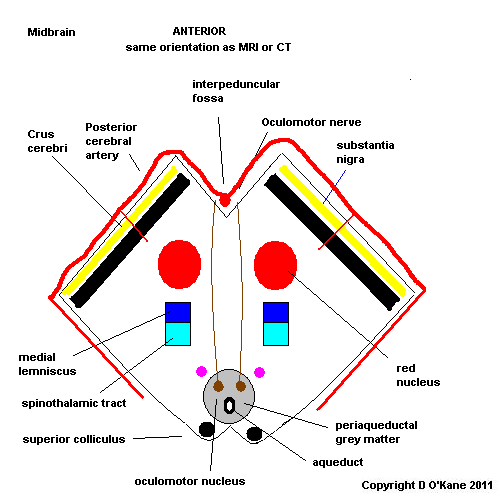
Inferior Colliculus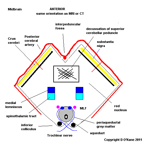
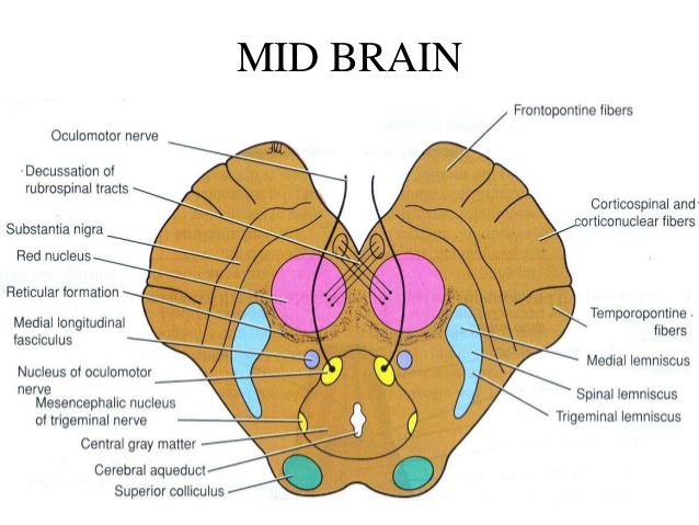
Midbrain at Super colliculus level
- Superior colliculus lies dorsally as a small hillock
- Cerebral aqueduct surrounded by periaqueductal grey matter
- Oculomotor nucleus on either side of midline in front of aqueduct. Supplies ipsilateral extraocular muscles and pupil. Fibres run forwards and exit anteriorly between cerebral peduncles
- Red nucleus lies on either side
- Substantia nigra lies between main brainstem and cerebral peduncles. Pigmented. Important in Parkinson's disease.
- Cerebral peduncles lie anteriorly and contain the corticonuclear fibres medially and the corticospinal fibres laterally
- Medial longitudinal fasciculus
- Medial lemniscus
- Dentatothalamic tract
- Medial Geniculate body lies laterally
- Reticular activating system
Midbrain at Inferior colliculus level
- Inferior colliculus lies dorsally
- IVth nerve nucleus is found at this level just on either side of midline
In the midline posteriorly lies the aqueduct carrying CSF from IIIrd to IVth ventricle. Surrounded by grey matter (cell bodies) called the periaqueductal grey matter which is involved in pain perception and stress responses. Midbrain also colourful as more anteriorly it has the red nucleus and the substantia nigra.
Vascular supply
- Vascular supply is from the basilar artery and the posterior cerebral artery. Infarction can be medial, laterally, rostral or caudal and can extend to thalamus and cortical areas supplied by PCA.