Learning objectives
- Learning
- Understand
- Integrate
- Reflect
|
Introduction
- Bleeds are usually unilateral and hemispheric.
- Some may need urgent neurosurgical input with clot evacuation for compression and/or shunting for acute obstructive hydrocephalus.
- It is a cause of acute usually non transient vertigo with other symptoms
Location
- Midline: lesions can damage deep cerebellar nuclei and pontine compression with related signs
- Lateral: Unilateral and Hemispheric (better prognostically) as less brainstem (pontine) compression. The more lateral the bleed the better
Causes
- Hypertensive rupture of small penetrating vessels
- Amyloid angiopathy, Cocaine
- AV malformations, Tumours with haemorrhage
- Anticoagulants / Coagulopathies
- Spinal surgery, Spontaneous intracranial hypotension
Clinical
- Abrupt onset of nausea, vertigo, headache, vomiting
- Ipsilateral cerebellar signs and VI and VII nerve lesions
- (Truncal) Ataxic gait unable to walk usually
- Coma and pupillary abnormalities suggest compression
- Neck stiffness and pain
- Coma, loss of corneal reflexes, pinpoint pupils
- Extensor plantars, Cheyne-stokes respiration
Differentials
- Labyrinthitis
- Alcohol - its not unique for a person with a cerebellar stroke to spend a night in the drunk tank or the drunk person to be admitted as stroke
- Drug toxicity - anticonvulsants
Poor outcome/Complications
- Haematoma > 3 cm
- Swelling and pressure on brainstem causing coma with increased pressure and reduced perfusion of brainstem
- Acute Obstructing Hydrocephalus usually at IVth ventricle
- Midline lesions
- Intraventricular extension of bleeding
- Low GCS
Investigations
- FBC, Coagulation Screen (INR if on warfarin), U&E, LFTs
- ECG and CXR if needed
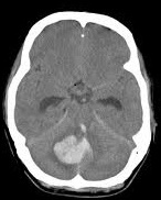
- Urgent CT head is usually diagnostic. Measure maximal diameter of bleed and look for early or developed hydrocephalus. Look for brainstem distortion and blood in ventricles.
- MRI may be performed if stable especially if there is suspect structural lesion
Management
- ABC and consider ITU if appropriate in rapidly deteriorating or comatose patient usually as a bridge to surgery so take neurosurgical advice. There can be profound benefits from timely surgery in those where brainstem compression or hydrocephalus is imminent or in progress. If patient is awake with a near normal GCS(14/15) and the bleed is less than 3 cm and no sign of hydrocephalus then these patients may be managed conservatively with an option to operate with any deterioration but best to inform neurosurgeons so if needed they are familiar with the case. The operative choices may be Ventriculostomy and drainage with significant haemorrhage and hydrocephalus or Suboccipital craniotomy with clot evacuation is indicated in patients with reduced LOC and clot usually > 3-4 cm in diameter. Consider Mannitol 1 g/kg acutely as a bridge to surgical treatment in those with evidence of increased pressure.
- Correct any coagulopathy e.g. Octaplex and IV Vitamin K or similar for Warfarin. Cautiously control any high BP. Discuss with neurosurgeons.
- Admit stroke Unit and manage as any haemorrhagic stroke with monitoring for deterioration if not for urgent surgery.
- Escalate in any change in condition. For those with significant coma, brainstem signs then end of life care may be indicated. Take expert advice.
- Do not forget the role of good Multidisciplinary stroke unit care and rehabilitation as these patients can often do well with time and support.