Themes: Stroke, Visual loss, Cardiac disease
| John is a 72 year old gentleman. He had an accident with his car this morning and clipped all the cars that were parked on the left side and he almost hit a cyclist. His wife who was with him feels he is "not right" since the evening before with chest discomfort which he has said is indigestion. He has been diabetic for 20 years and has hypertension and smokes 20/day. There is no weakness and no sensory loss. He feels too fatigued to walk but on assessment you note that he has a hemianopia. |  |
|---|
1. He has a CT head scan. Look at it closely.

2. What blood vessel has been blocked to cause his hemianopia
He has a Right Posterior cerebral artery infarct which would mean he cannot see to his left
3. Is this an anterior or posterior circulation stroke
In most patients the PCA is formed by the splitting of the basilar artery and is supplied by blood from the vertebrobasilar system so this is a Posterior circulation infarct (POCI).
4. What lobe of the brain has been affected
The right occipital lobe which deals with vision from the left
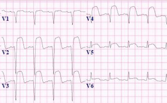
As part of his work up in the emergency department an ECG is done. He denies any chest pain but had some new chest discomfort the evening before. He admits to being an occasional smoker and stopped taking a statin for hypercholesterolaemia
5. Look at the ECG show

6. What does the ECG show
7. His troponin is markedly raised. He has a soft pansystolic murmur and a 3rd heart sound and some bibasal fine inspiratory crackles. What is the next test that should be performed. It is now over 24 hrs since he had chest symptoms.
| He needs a transthoracic echocardiogram as the concern would be that he has an anterior STEMI with mural thrombus. It would be useful to assess his LV function, exclude mural thrombus and also to assess LA size and valves. This should be done soon. The stroke physician calls the cardiologists. They want an echocardiogram and they will do an angiogram on the next listy. The echocardiogram shows severe LV dyskinesia and an ejection fraction of 20%. There is some mild MR but the LA looks normal. He is given dual antiplatelet therapy. He is started on ramipril and a mild diuretic. |
|---|
8. He has a coronary angiogram and stent to his LAD and is placed on Dual antiplatelet therapy. He does well and goes home day 7. His hemianopia has improved. He has no signs of cardiac failure. He tells you that he will never smoke again. He will see his GP about diabetic monitoring which he had missed. he is going to do some cardiac rehabilitation. He asks about driving.
| Note: The plan is to keep the website free through donations and advertisers that do not present any conflicts of interest. I am keen to advertise courses and conferences. If you have found the site useful or have any constructive comments please write to me at drokane (at) gmail.com. I keep a list of patrons to whom I am indebted who have contributed. If you would like to advertise a course or conference then please contact me directly for costs and to discuss a sponsored link from this site. |