Learning objectives
|
Introduction
- This is an important cause of stroke predominantly of younger adults.
- Always ask about unusual neck position or turning or trauma in young strokes
Vertebral artery anatomy
- The VA is described as having 4 anatomical segments (V1-V4)
- V1 From its origin at the subclavian artery to its entry into the transverse foramen of C6
- V2 From the transverse foramen of C6 cranially to where it exits the C1 transverse foramen to become V3
- V3:wraps around C1 and ascends into the foramen magnum
- V4: intracranial portion extends from the foramen magnum where it pierces the dura and joins the contralateral VA to form the basilar artery, which perfuses the brainstem, cerebellum, and occipital lobes.

| Extracranial dissection is more common than intracranial, but the V2 segment is the least common of the extracranial segments to dissect because it remains relatively fixed within the transverse foramina along its course. The first and third segments are highly mobile and, thus, are more frequently involved in dissection |
Aetiology
- Consider and screen for it in any younger person (under 50) with a posterior circulation stroke particularly if there is neck pain or where there is no alternative aetiology found.
- Dissection leads to endothelial luminal clot formation which can embolise.
- The clot can also cause occlusion of the vertebral or embolise into basilar and then posterior cerebral arteries in branches such as those to the cerebellum. Brainstem, thalamic, occipital and cerebellar infarcts may be seen.
- Thromboembolism originating at vertebral level may cause infarcts above the basilar artery on either side of the posterior circulation (occipital cortex and thalamus).
Risks factors
- Ehlers Danlos Type IV
- Fibromuscular dysplasia
- Neck trauma e.g. hairdressers, reversing car looking over shoulder.
- Peripartum period
Aetiology
- Dissection and vertebral occlusion can cause an ipsilateral lateral medullary syndrome
- Horner's syndrome due to disruption of the tracts in the lateral medulla if dissection causes PICA infarct
- There is a small risk of intracranial dissections bleeding causing SAH (thunderclap headache).
- Usually affects V3 or less so the V2 segments of the vertebral artery
| Causes of Dissection seen recently | |||
|---|---|---|---|
 |
 |
 |
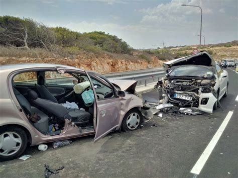 |
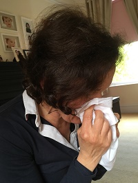
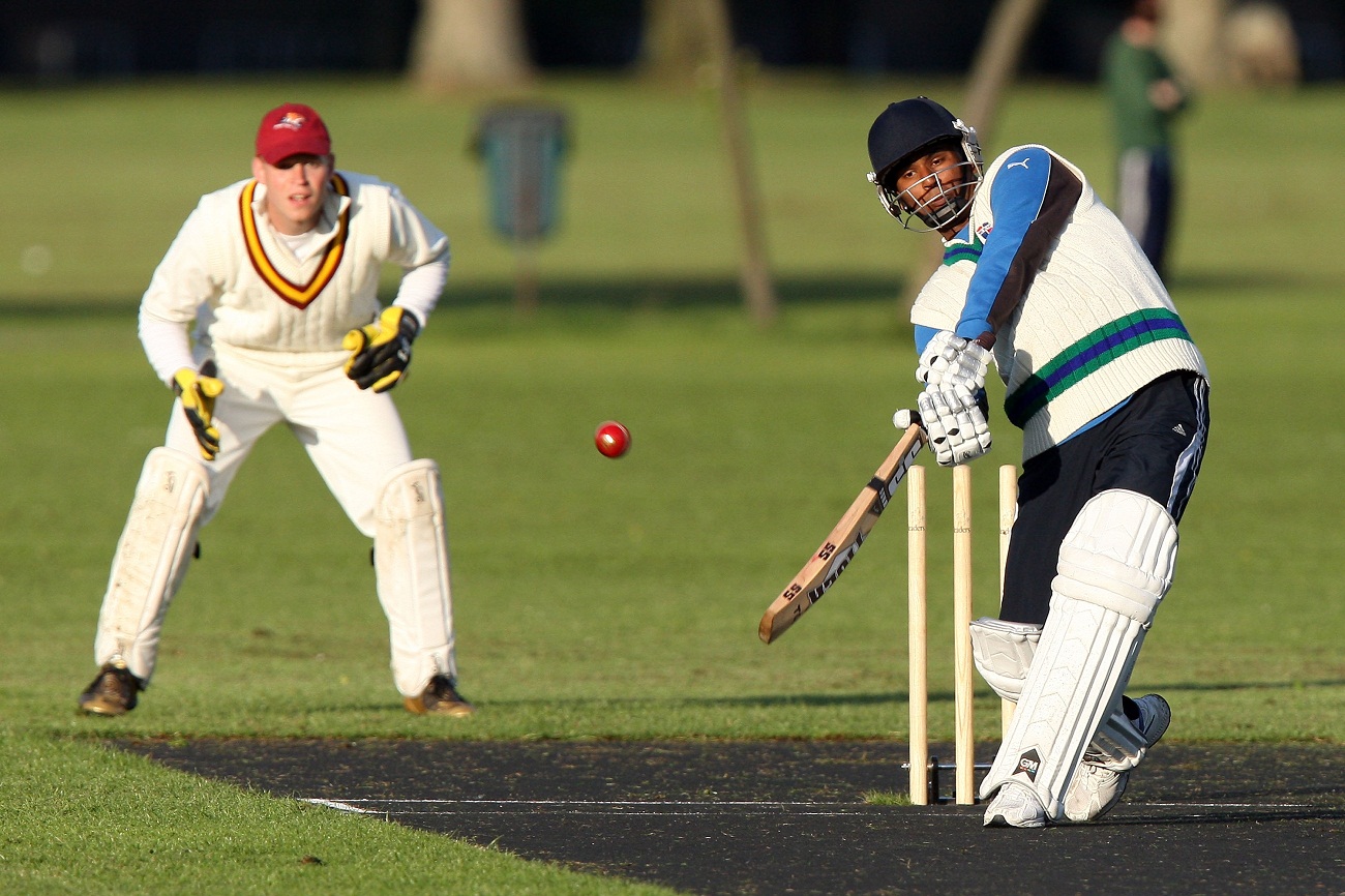
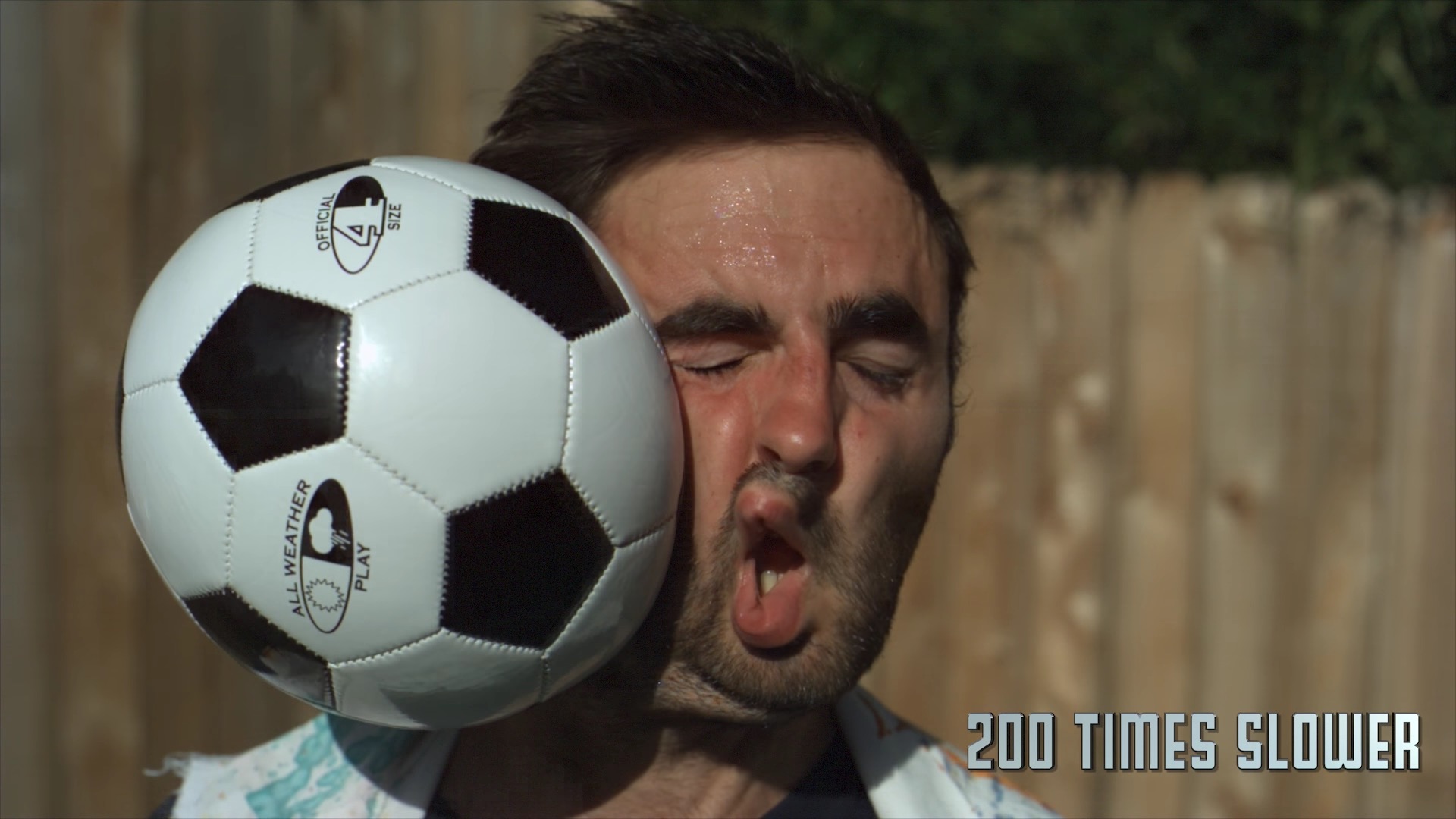
| A sneeze or other violent neck movement. | Sports such as cricket and rowing. | Blow to the head causing torsional forces - hit with a football. | Following a road traffic accident. |
| Some possible associations with dissection |
|---|
|
| The issue with chiropracter manipulation is that it may be done to ease neck pain which may have been due to a dissection. It may just be association rather than causation for some. |
Clinical
| Presentations of Vertebral Dissection |
|---|
|
Differential
- Cervical spondylosis and upper limb radiculopathy
Investigations
- MRI T1 fat-suppression technique (wall haematoma) to shows crescent shaped intramural clot within the relevant vertebral artery.
- MRI DWI may show infarction anywhere in the posterior circulation due to showering of emboli. This would include the brainstem, either cerebellar hemisphere or medial temporal lobe or thalamus or occipital cortex.
- CTA/MRA are also done to show the level of any blockage and to demonstrate infarction. Classically the dissection shows a narrowed lumen surrounded by mural thickening with mild tapering rather than an abrupt stenosis seen with embolism or thrombosis in situ.
Management
- ABC and acute thrombolysis/thrombectomy is indicated. Vessel rupture is very rare indeed and dissection is not a bar to reperfusion therapies. However pure POCI are often more diffiicult to reliably diagnose and are often but not always outside therapy window when diagnosis considered.
- Most extracranial VADs carry a good prognosis, heal spontaneously with time, and rarely require surgical intervention unless complicated by other factors, such as intracranial extension
- The patient may simply present with posterior circulation stroke symptoms which may be mild and neck pain. Management is the same as for carotid dissection.
- The fact that it has not been shown that anticoagulation is better than anti-platelet therapy makes life simpler when the imaging is equivocal, or the presentation is late one can simply treat as ischaemic stroke with either aspirin or clopidogrel or even dual therapy for a short period that theoretically allows the interrupted possibly prothrombotic intima to repair and re-endothelialise.
- Carotid or vertebral dissection without evidence or suspicion of SAH is not a contraindication to thrombolysis.
- Risk of recurrence seems low especially if the provoking mechanism is avoided.
Further reading and references
- Carotid and vertebral artery dissections . Arnold M, Bousser†MG. Practical Neurology, 2005, 5, 100–109
- Vertebral Artery Dissection Presenting Findings and Predictors of Outcome. Stroke. 2006;37:2499-2503
| Note: The plan is to keep the website free through donations and advertisers that do not present any conflicts of interest. I am keen to advertise courses and conferences. If you have found the site useful or have any constructive comments please write to me at drokane (at) gmail.com. I keep a list of patrons to whom I am indebted who have contributed. If you would like to advertise a course or conference then please contact me directly for costs and to discuss a sponsored link from this site. |